The Nikon Small World Photomicrography Competition lets us see beyond the capabilities of our unaided eyes. Almost 2000 entries from 70 countries vied for recognition in the 37th annual contest, which celebrates photography through a microscope. Images two through 21 showcase the contest's winners in order, and are followed by a selection of other outstanding works. Scientists and photographers turned their attention on a wide range of subjects, both living and man-made, from lacewing larva to charged couple devices, sometimes magnifying them over 2000 times their original size. --
Lane Turner
Lane Turner
 |
| Dr. Donna Stolz of the University of Pittsburgh assembled a wreath collage of mammalian cells stained for various proteins and organelles magnified from 220x to 2000x. (Dr. Donna Stolz) |
 |
| A confocal image of a reconstruction of a fruit fly (Drosophila sp.) nervous system by Dr. Jana Boerner of Florida Atlantic University in Boca Raton, Fla. (Dr. Jana Boerner) |
 |
| Jonathan Franks of the University of Pittsburgh used the confocal method with autofluorescence to capture algae biofilm. (Jonathan Franks) |
 |
| An antique mount of a blowfly (Calliphoridae) proboscis was photographed by Dr. Davis Linstead of Kent, UK with differential interference contrast. (Dr. David Linstead) |
 |
| The double compound eyes of a male St. Mark's fly (Bibio marci) photographed with reflected (episcopic) diffuse illumination by Dr. David Maitland of Feltwell, UK. (Dr. David Maitland) |
 |
| The egg of a red admiral butterfly (Vanessa atalanta) in stinging nettle (Urtica dioica) trichomes photographed by David Millard of Austin, Texas with diffuse incident illumination. (David Millard) |
 |
| Marek Mis of Suwalki, Poland used polarized light to photograph green algae (Spirogyra sp.) filaments. (Marek Mis) |
 |
| The eyes (anterior lateral and median) of a jumping spider photographed in reflected light by Walter Piorkowski of South Beloit, Illinois. (Walter Piorkowski) |
 |
| A confocal image of a charge coupled device (CCD) sensor, direct surface view, magnified 1000 times by Kevin Smith of MetPrep Ltd. in Warwickshire, UK. (Kevin Smith) |
 |
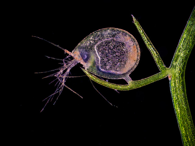
| A water flea (Daphnia sp.) and green algae (Volvox sp.) captured with darkfield and flash by Dr. Ralf Wagner of Dusseldorf, Germany. (Dr. Ralf Wagner) |
 |
| The mouth of a common fly photographed with fiber optic illumination by Dr. Havi Sarfaty of the Israeli Veterinary Association. (Dr. Havi Sarfaty) |


